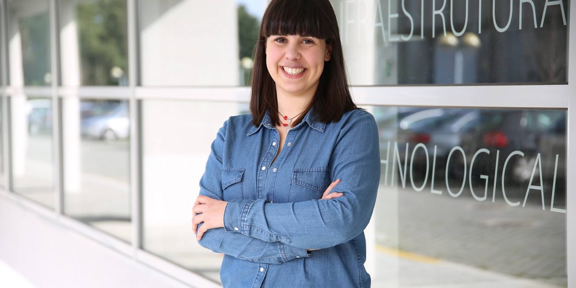
By Sara Oliveira, Researcher of INESC TEC’s Centre for Telecommunications and Multimedia (CTM)
It’s October, the month that is dedicated to the breast cancer awareness and to the importance of prevention and early diagnosis of the disease For this reason, when I was invited to write for BIP, I considered it to be a great opportunity to present you my PhD project.
I am a member of the VCMI (Visual Computing and Machine Intelligence) research group, which integrates CTM, led by Professor Jaime Cardoso and, in particular, of the Breast Research Group, whose research line is focused on breast cancer in close connection with the breast unit of the Champalimaud Foundation. I arrived at INESC TEC in 2016 as a research grantee of the BCCT.plan, a project that focuses on the development of a 3D custom surgical planning tool for the breast cancer conservative surgery. In the meantime, in 2018, I started my PhD at FEUP, being supervised by researcher and Professor Hélder Oliveira, and I changed my work’s direction a little bit. I moved on from the 3D world to the two dimensions and I decided to explore approaches of medical image analysis and processing, known as “radiomics” and “pathomics”. And what is this? Basically, they are methods for converting images into a set of data, going beyond those that are visible to the “naked eye”, and exploiting their potential in order to broaden the tumour characterisation.
The use of medical image to support the breast cancer diagnosis has been part of clinical practice for a while now, but the full characterisation of the tumour is still a challenging task due to the partiality of the information that can be obtained with each imaging modality. And this is where we found a research opportunity: why not combine the information that each modality contains and strengthen the support models for clinical decision making?!
From the screening/diagnosis of the disease to its treatment, several radiological exams are performed (mammography, magnetic resonance, echography) and a diverse set of information is gathered. In addition to this, after the breast cancer diagnosis, it is important to have even more detailed information about the characteristics of the tumour. This way, a biopsy is performed in which a tissue sample is obtained and then, after being prepared, it results in a (histopathological) image. In the end, each case has an associated set of images: the raw material for our work. Therefore, the goal is to explore imaging processing techniques and, in particular, deep learning techniques for an effective and accurate combination of all these images (particularly those of the magnetic resonance and the histopathological ones) for the classification of the breast cancer subtype.
And what are the research challenges? In fact, this problem becomes particularly challenging mainly due to three factors: the subtlety of lesions, which in many cases makes them more difficult to detect; the high resolution of images, especially the histopathology ones, which require larger models, thus needing more data that sometimes is rare to find; and, finally, the heterogeneity of the type of images, which implies the use of different approaches and methods for their analysis. However, this is what research is for: so we can’t let ourselves be intimidated by the challenges and to search for ways to solve them. And if I’m allowed to say, I hope they inspire us to think outside the box!


 News, current topics, curiosities and so much more about INESC TEC and its community!