INESC TEC Science Bits – Episode 38
Link to the episode (Portuguese only)
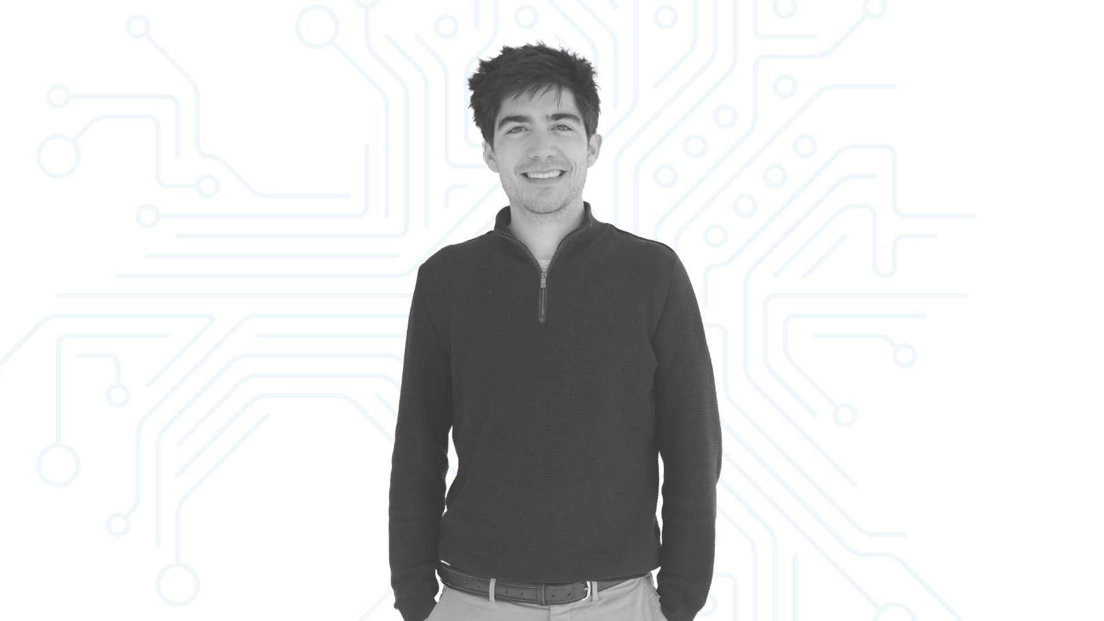
Guest speaker: João Pedrosa, researcher at INESC TEC
Keywords: Artificial Intelligence, Medical Image Analysis, Radiology, Lung Cancer, Explainability

We kick off 2024 by decoding, bit by bit, AI-supported medical diagnosis. But before that, shall we take a quick trip back, to the 19th century? It’s November 8, 1895, and we’re inside Wilhelm Conrad Röntgen’s laboratory – the first Nobel Prize winner in Physics, also known as the “father of Radiology”. Röntgen discovered – supposedly, by accident – the X-rays, while studying cathode rays. Through a series of experiments, this German physicist realised that X-rays could penetrate the skin and muscle, but not the bones, and that they can be photographed. This innovation soon started to be used in medical diagnosis – until the present day.
Nearly 130 years later, the commonly known X-ray remains a frequently used examination technique. In some hospitals, including Portuguese hospitals, it is now combined with Artificial Intelligence technologies. How? That’s what we’re going to discover in this conversation with João Pedrosa, researcher at INESC TEC, and invited assistant professor at the Faculty of Engineering of the University of Porto.


 News, current topics, curiosities and so much more about INESC TEC and its community!