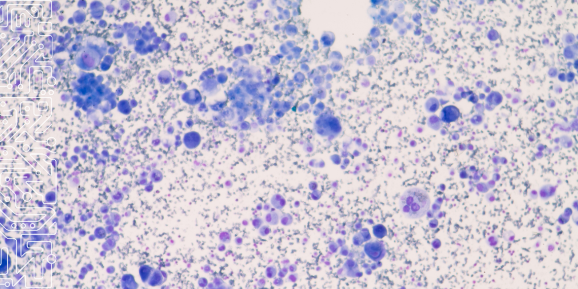
The cell nucleus contains crucial information that supports the diagnosis of diseases, considering the prediction of their progression and the planning of appropriate and effective treatments – namely in cancer diagnoses.
The analysis of these nuclei is already performed, either visually, by pathologists, or in an automated way, using image processing algorithms. Now, thorough research into the methods of using context and attention in cell nuclei segmentation and classification tasks – carried out by João Nunes, a researcher at INESC TEC, and Diana Montezuma Felizardo, an anatomopathologist and researcher at IMP Diagnostics -, introduces a deeper approach.
“A Survey on Cell Nuclei Instance Segmentation and Classification: Leveraging Context and Attention”, is the name of the article published in the journal Medical Image Analysis, which integrates the TOP 10 publications in computer vision and pattern recognition and explores the importance of analysing the context of the cell nucleus to achieve more precise segmentation.
With AI as a major topic nowadays – regarding the Nobel Prizes in Physics and Chemistry – this research takes another step in the application of machine learning to other areas, like medicine.
“The nuclei are the cellular structures that contain the genetic material (DNA), responsible for controlling the activities of the cell,” said Diana Montezuma, stressing that “the analysis of the nuclei is crucial, as changes in their shape, size and structure can indicate the presence of diseases, namely cancer.”
João Nunes explained that this work “addresses the different mechanisms of attention and context for the detection of cell nuclei as a way to overcome the main difficulties of the task, including overlapping or intersecting cells, irrelevant information in the background of the image, cells arranged in a disorganised way and elements like blurred regions, air bubbles, etc. Among the benefits is the ability to suppress noisy or irrelevant information, as well as the potential for models to pay more attention to the boundary between nuclei, thus enabling a better separation of instances.”
The authors highlighted that the manual segmentation of these images is a complex and time-consuming task, addressing the use of automatic core segmentation and classification algorithms to reduce the workload of pathologists and improve the extraction of clinically interpretable features. Meanwhile, current algorithms face difficulties in dealing with variabilities in the morphological and chromatic characteristics of the nuclei.
To address these challenges, the researchers focused on improving algorithms by adding “attention” and “context” mechanisms. By ensuring that the algorithm “pays attention” to what surrounds cells and their most crucial features, the accuracy of segmentation and classification improves. This helps doctors make faster and more reliable decisions, optimising time and resources in the treatment of serious diseases like cancer.
“The improvement of machine learning techniques for detecting and classifying the nuclei may have several clinical implications,” said the researcher. “A practical example is that the appearance and shape of tumor cell nuclei is used to determine the degree of some types of cancer, namely breast cancer. A better segmentation and classification of the nuclei will allow a better characterisation of the tumor cells (and other cells of the tumor microenvironment), thus contributing to a greater diagnostic and prognostic acuity of the patients.”
In the research, INESC TEC was responsible for reviewing the existing algorithms, as well as for the design and development of the experiments, while the IMP Diagnostics team was responsible for the clinical dimension of the study – namely framing the importance of the detection and segmentation of cell nuclei for the diagnosis and prognosis of tumors, as well as other pathologies.
The article is available here.
The researcher mentioned in this news piece are associated with INESC TEC.


 News, current topics, curiosities and so much more about INESC TEC and its community!